
Matte High Definition Full Color

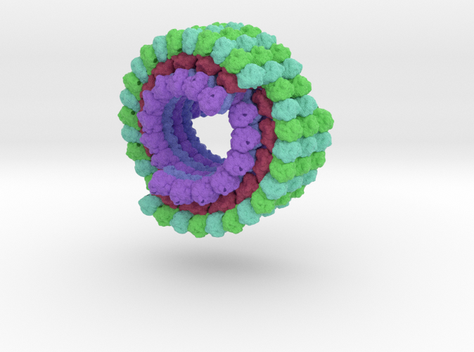
Microtubule 3J2U
Made by
Print With Shapeways
 Choose Your Size
 Choose Your Material
 Choose your color and finish
$305.84
Have a question about this product?
contact the designerYou must be logged in and verified to contact the designer.
Product Description
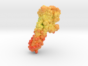
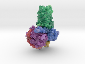
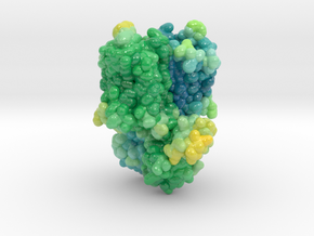
To elucidate the structural basis of the mechanism of microtubule depolymerization by kinesin-13s, we analyzed complexes of tubulin and the Drosophila melanogaster kinesin-13 KLP10A by electron microscopy (EM) and fluorescence polarization microscopy. We report a nanometer-resolution (1.1 nm) cryo-EM three-dimensional structure of the KLP10A head domain (KLP10AHD) bound to curved tubulin. We found that binding of KLP10AHD induces a distinct tubulin configuration with displacement (shear) between tubulin subunits in addition to curvature. In this configuration, the kinesin-binding site differs from that in straight tubulin, providing an explanation for the distinct interaction modes of kinesin-13s with the microtubule lattice or its ends. The KLP10AHD-tubulin interface comprises three areas of interaction, suggesting a crossbow-type tubulin-bending mechanism. These areas include the kinesin-13 family conserved KVD residues, and as predicted from the crossbow model, mutating these residues changes the orientation and mobility of KLP10AHDs interacting with the microtubule.
Details
What's in the box:
sml_Microtubule-3j2u_max-d025_x75-6cm_E24 (repaire
Dimensions:
Success Rate:
First To try.
What's this?
Rating:
Mature audiences only.
