Printed in sandstone


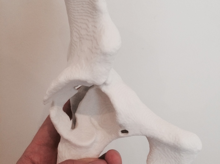
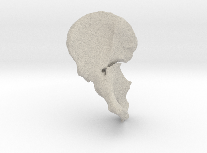
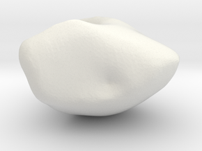
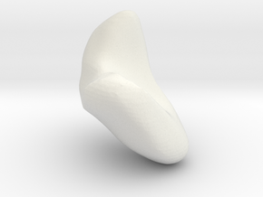
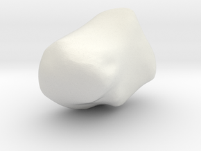
Acetabular Fracture
Made by
Print With Shapeways
 Choose Your Material
 Choose your color and finish
$156.02
Have a question about this product?
contact the designerYou must be logged in and verified to contact the designer.
Product Description
Lifesize model of a fractured acetabulum created from CT data.
Orthopaedic pre-operative planning often uses 3D reconstructions of CT data. In this case I took raw data (0.6mm spirally acquired slices) and created this exquisitely accurate segmentation of the left iliac blade, which demonstrates a complex acetabular blow-out fracture.
I have always been fascinated by anatomy. By day I'm a radiologist, reporting X-ray, CT and MRI scans on a wide variety of patients. I get to work with state-of-the-art scanners and front-line diagnostic imaging in a busy hospital - but by night I take my passion home and create accurate and detailed anatomical models. I use high resolution scan data and interesting cases to bring anatomy and pathology to life. I'm still honing my skills, streamlining the workflow and improving, but with the advent of 3D printing I can now bring my dreams into the real world.
See more at www.drhughharvey.com
Details
What's in the box:
Acetabular Fracture
Dimensions:
Success Rate:
First To try.
What's this?
Rating:
Mature audiences only.