Glossy Full Color Sandstone

Oxygenated Hemoglobin (Hb) 1HHO
Made by
Print With Shapeways
 Choose Your Size
 Choose Your Material
 Choose your color and finish
$156.92
Have a question about this product?
contact the designerYou must be logged in and verified to contact the designer.
Product Description
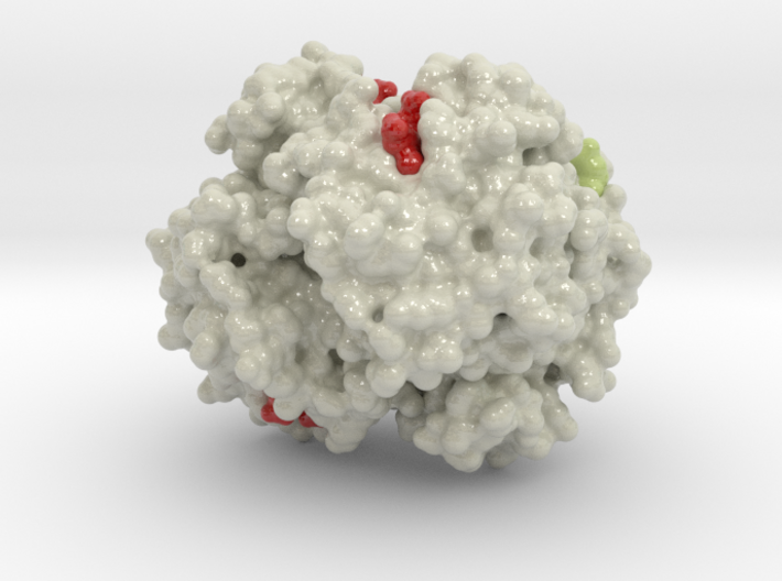
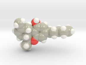
3D printed protein model of Oxygenated Hemoglobin Hb created from PDB ID: 1HHO.
Model Description
3D reproduction of Oxygenated Hemoglobin Hb, 18 Million times larger than diameter actual protein. Colored in red are the protein’s HEME Groups, disk-like structures with an Iron atom core. It is to this Iron atom core that binds O2 molecules (Blue) bind. This protein model is designed to visual the mutation properties of Sickle Cell Anemia and demonstrate binding of two sickled hemoglobins together. Val 6 of each Beta chain (the residue that mutates in Sickle Cell Anemia) is colored light yellow and the two residues that Phe85 and Leu88 (light and dark grey). Protein model base available here
Protein Description
Oxygenated Hemoglobin (Hb) is the protein from inside red blood cells that transporters small molecules like Oxygen (O2) throughout the body. It is a specialized protein exhibiting complex molecular behaviors. Hemoglobin is an important protein to life, specialized at binding and carrying one of the most fundamental molecules for life on our planet, Oxygen. Despite hemoglobin being millions of times smaller than anything we can see with our eyes, we can experience hemoglobin in our everyday life. Hemoglobin gives our blood its red color. When our cheeks become flush with embarrassment, it's hemoglobin that makes them look rosy. The immediate consequence of taking a breath is for hemoglobin to bind oxygen and change shape. While it may be a tiny, we can draw a deeper understanding of the world around us through this little thing.
Details
What's in the box:
mdm_hemogloibin_3DPrint_max_B11

Dimensions:
Success Rate:
First To try.
What's this?
Rating:
Mature audiences only.
