Glossy Full Color Sandstone

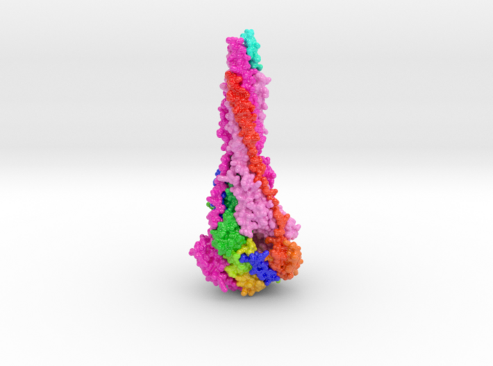
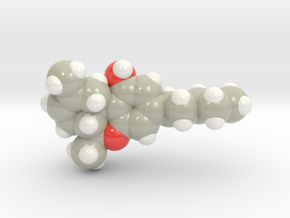
RSV Fusion Glycoprotein Postfusion 3RRR
Made by
Print With Shapeways
 Choose Your Size
 Choose Your Material
 Choose your color and finish
$68.83
Have a question about this product?
contact the designerYou must be logged in and verified to contact the designer.
Product Description
Model Description
This 3D printed protein model of RSV Fusion Glycoprotein colored shades of magenta to visualize the viral binding protein's three fusion (F) chains. One chain is colored as a blue to red from N to C Terminal. This model was created from PDB ID: 3RRR and paired with RSV Fusion Glycoprotein Prefusion 3D printed protein model.
Protein Description
Respiratory syncytial virus (RSV) invades host cells via a type I fusion (F) glycoprotein that undergoes dramatic structural rearrangements during the fusion process. Neutralizing monoclonal antibodies, such as 101F, palivizumab, and motavizumab, target two major antigenic sites on the RSV F glycoprotein. The structures of these sites as peptide complexes with motavizumab and 101F have been previously determined, but a structure for the trimeric RSV F glycoprotein ectodomain has remained elusive. To address this issue, we undertook structural and biophysical studies on stable ectodomain constructs. Here, we present the 2.8-Å crystal structure of the trimeric RSV F ectodomain in its postfusion conformation. The structure revealed that the 101F and motavizumab epitopes are present in the postfusion state and that their conformations are similar to those observed in the antibody-bound peptide structures. Both antibodies bound the postfusion F glycoprotein with high affinity in surface plasmon resonance experiments. Modeling of the antibodies bound to the F glycoprotein predicts that the 101F epitope is larger than the linear peptide and restricted to a single protomer in the trimer, whereas motavizumab likely contacts residues on two protomers, indicating a quaternary epitope. Mechanistically, these results suggest that 101F and motavizumab can bind to multiple conformations of the fusion glycoprotein and can neutralize late in the entry process. The structural preservation of neutralizing epitopes in the postfusion state suggests that this confirmation can elicit neutralizing antibodies and serve as a useful vaccine antigen.
https://www.rcsb.org/structure/3RRR
Details
What's in the box:
sml_prefusion_5tdl_beta_x75-5cm_ml_vF14

Dimensions:
Success Rate:
First To try.
What's this?
Rating:
Mature audiences only.
