Smooth Full Color Nylon 12 (MJF)

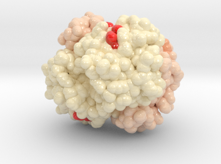
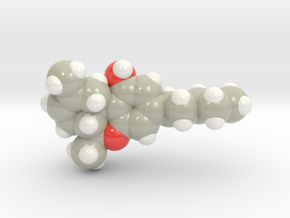
Oxygenated Hemoglobin 1HHO
Made by
Print With Shapeways
 Choose Your Size
 Choose Your Material
 Choose your color and finish
$122.43
Have a question about this product?
contact the designerYou must be logged in and verified to contact the designer.
Product Description
The structure of human oxyhaemoglobin was determined by single crystal X-ray analysis at 2.1 A resolution. Data were collected on an Arndt-Wonacott camera at -2 degrees C. The structure was refined to an R factor of 0.223 by the Jack-Levitt method, starting from Baldwin's model of human carbon monoxide haemoglobin. The active sites in the alpha and beta subunit are distinct. The iron atoms are 0.16(8) A and 0.00(8) A from the mean plane of the porphyrin carbons and nitrogens (0.12(8) A and -0.11(8) A from the mean plane of the porphyrin nitrogens) in the alpha and beta subunit, respectively, in correlation with the orientation of HisF8 relative to the porphyrin nitrogens. The haem group appears to be nearly planar in the alpha subunit but ruffled in the beta subunit. The Fe-O(1)-O(2) angles are 153(7) degrees and 159(12) degrees in the alpha and beta subunit, respectively. The oxygen molecule forms a hydrogen bond to N epsilon of HisE7 in the alpha, but either none or a weak one in the beta subunit. The following bond lengths were found: Fe-N epsilon (HisF8) = 1.94(9) A (alpha) and 2.07(9) A (beta); Fe-O(1) = 1.66(8) A (alpha) and 1.87(13) A (beta); Fe-Nporph (mean = 1.99(5) A (alpha) and 1.96(6) A (beta). These dimensions agree with the values obtained in oxymyoglobin and model compounds. The C-terminal residues, ArgHC3(141 alpha) and HisHC3(146 beta), are relatively delocalized, and their positions do not enable them to form the intersubunit salt bridges in which they are involved in deoxyhaemoglobin. The penultimate tyrosine residues, TyrHC2 140 alpha and 145 beta, are relatively localized and maintain the hydrogen bonds to the carbonyl oxygens of ValFG5 (93 alpha and 98 beta), with only minor variations compared to their geometry in deoxyhaemoglobin. TyrHC2(145 beta), however, alternates between a major and a minor site, in conjunction with CysF9(93 beta), both sharing the internal pocket between the F and H helices while in the major conformation. This suggests that the role of the penultimate tyrosines in the allosteric mechanism may differ from that previously proposed by Perutz. The overall quaternary structure of oxyhaemoglobin is identical, within experimental error, to that of carbon monoxide haemoglobin, and thus confirms the applicability of the allosteric mechanisms proposed by Perutz and Baldwin & Chothia to the process of oxygen binding.
Details
What's in the box:
exsml_Hb_HD_1HHO_ml_vA13_x42_3cm

Dimensions:
Success Rate:
First To try.
What's this?
Rating:
Mature audiences only.
