Glossy Full Color Sandstone

Product Description
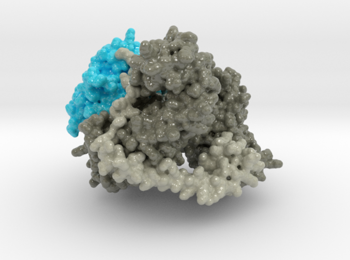
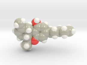
3D printed protein model of Human NatA created from PDB ID: 6C95.
Model Description
This 3D printed protein model of Human NatA is visualized as a volumetric surface and colored to distinguish how multiple chains interact. Chain B is colored cyan and residue 100 in red. Chains A and C colored light and dark grey.
Protein Description
Co-translational N-terminal protein acetylation regulates many protein functions including degradation, folding, interprotein interactions, and targeting. Human NatA (hNatA), one of six conserved metazoan N-terminal acetyltransferases, contains Naa10 catalytic and Naa15 auxiliary subunits, and associates with the intrinsically disordered Huntingtin yeast two-hybrid protein K (HYPK). We report on the crystal structures of hNatA and hNatA/HYPK, and associated biochemical and enzymatic analyses. We demonstrate that hNatA contains unique features: a stabilizing inositol hexaphosphate (IP6) molecule and a metazoan-specific Naa15 domain that mediates high-affinity HYPK binding. We find that HYPK harbors intrinsic hNatA-specific inhibitory activity through a bipartite structure: a ubiquitin-associated domain that binds a hNaa15 metazoan-specific region and an N-terminal loop-helix region that distorts the hNaa10 active site. We show that HYPK binding blocks hNaa50 targeting to hNatA, likely limiting Naa50 ribosome localization in vivo. These studies provide a model for metazoan NAT activity and HYPK regulation of N-terminal acetylation.
Details
What's in the box:
mdm_human_natA_6C95_ml_v1_x83

Dimensions:
Success Rate:
First To try.
What's this?
Rating:
Mature audiences only.
