Viral art

Designs by DavidBhella
The models on offer here were generated by imaging viruses in the electron microscope. The resulting micrographs were computationally processed to extract the three-dimensional structure at intermediate resolution.

We found 12 products by DavidBhella

by DavidBhella
This is a three-dimensional reconstruction of a Herpes Simplex Virus capsid, solved by cryo-electron microscopy. It is therefore an exact scale model of the virus capsid (approximately 400,000 x).
 
(5)  
  •  
From: $24.33
 
by DavidBhella
This is a three-dimensional reconstruction of a Herpes Simplex Virus capsid, solved by cryo-electron microscopy. It is therefore an exact scale model of the virus capsid (approximately 800,000 x)
 
(0)  
  •  
From: $120.20
 
by DavidBhella
This is a three-dimensional reconstruction of Feline Calicivirus solved by cryo-electron microscopy. It is therefore an exact scale model of the virus (approximately 2,000,000 x). This structure was reported in "The cryo-electron microscopy structure of feline calicivirus bound to junctional adhesion molecule A at 9-angstrom resolution reveals receptor-induced flexibility and two distinct conformational changes in the capsid protein VP1" Bhella D, Goodfellow IG. J Virol. 2011 Nov;85(21):11381-90
 
(0)  
  •  
From: $98.64
 
by DavidBhella
This is a three-dimensional reconstruction of Feline Calicivirus bound to its cellular receptor Junctional Adhesion Molecule A, solved by cryo-electron microscopy. It is therefore an exact scale model of the virus receptor complex (approximately 2,000,000 x). Receptor binding induces a conformational change in the capsid, this model shows the structure before the conformational change. This structure was reported in "The cryo-electron microscopy structure of feline calicivirus bound to junctional adhesion molecule A at 9-angstrom resolution reveals receptor-induced flexibility and two distinct conformational changes in the capsid protein VP1" Bhella D, Goodfellow IG. J Virol. 2011 Nov;85(21):11381-90
 
(0)  
  •  
From: $187.45
 
by DavidBhella
This is a three-dimensional reconstruction of Feline Calicivirus bound to its cellular receptor Junctional Adhesion Molecule A, solved by cryo-electron microscopy. It is therefore an exact scale model of the virus receptor complex (approximately 2,000,000 x). Receptor binding induces a conformational change in the capsid, this model shows the structure after the conformational change. This structure was reported in "The cryo-electron microscopy structure of feline calicivirus bound to junctional adhesion molecule A at 9-angstrom resolution reveals receptor-induced flexibility and two distinct conformational changes in the capsid protein VP1" Bhella D, Goodfellow IG. J Virol. 2011 Nov;85(21):11381-90
 
(0)  
  •  
From: $188.14
 
by DavidBhella
This is a three-dimensional reconstruction of Feline Calicivirus bound to its cellular receptor Junctional Adhesion Molecule A, solved by cryo-electron microscopy. It is therefore an exact scale model of the virus receptor complex (approximately 1,000,000 x). Receptor binding induces a conformational change in the capsid, this model shows the structure before the conformational change. This structure was reported in "The cryo-electron microscopy structure of feline calicivirus bound to junctional adhesion molecule A at 9-angstrom resolution reveals receptor-induced flexibility and two distinct conformational changes in the capsid protein VP1" Bhella D, Goodfellow IG. J Virol. 2011 Nov;85(21):11381-90
 
(0)  
  •  
From: $42.76
 
by DavidBhella
This is a three-dimensional reconstruction of Feline Calicivirus solved by cryo-electron microscopy. It is therefore an exact scale model of the virus (approximately 1,000,000 x). This structure was reported in "The cryo-electron microscopy structure of feline calicivirus bound to junctional adhesion molecule A at 9-angstrom resolution reveals receptor-induced flexibility and two distinct conformational changes in the capsid protein VP1" Bhella D, Goodfellow IG. J Virol. 2011 Nov;85(21):11381-90
 
(0)  
  •  
From: $27.23
 
by DavidBhella
This is a three-dimensional reconstruction of Feline Calicivirus bound to its cellular receptor Junctional Adhesion Molecule A, solved by cryo-electron microscopy. It is therefore an exact scale model of the virus receptor complex (approximately 1,000,000 x). Receptor binding induces a conformational change in the capsid, this model shows the structure after the conformational change. This structure was reported in "The cryo-electron microscopy structure of feline calicivirus bound to junctional adhesion molecule A at 9-angstrom resolution reveals receptor-induced flexibility and two distinct conformational changes in the capsid protein VP1" Bhella D, Goodfellow IG. J Virol. 2011 Nov;85(21):11381-90
 
(0)  
  •  
From: $42.85
 
by DavidBhella
This is a three-dimensional reconstruction of a Herpes Simplex Virus capsid, solved by cryo-electron microscopy. It is therefore an exact scale model of the virus capsid (approximately 1,000,000 x).
 
(0)  
  •  
From: $200.85
 
by DavidBhella
This is a three-dimensional reconstruction of a Herpes Simplex Virus capsid, solved by cryo-electron microscopy. It is therefore an exact scale model of the virus capsid (approximately 2,000,000 x). This is insanely expensive, please contact me if you want me to make it available in anything other tham WSF (if you are very very wealthy!)
 
(0)  
  •  
From: $1,511.44
 
by DavidBhella
This is a 3D reconstruction of the bacteriophage T7, solved by cryo-electron microscopy and 3D image processing. This model (which is not from my own research) is available as part of a schools project in partnership with the Society of General Microbiology. This model was produced from a reconstruction deposited in the EM database - EMD 1164 and published: Agirrezabala X, Martin-Benito J, Caston JR, Miranda R, Valpuesta JM, Carrascosa JL: Maturation of phage T7 involves structural modification of both shell and inner core components. EMBO J. (2005) 24, pp. 3820-3829 [PubMed 16211007]
 
(0)  
  •  
From: $35.78
 
by DavidBhella
This is a 3D reconstruction of the bacteriophage T7, solved by cryo-electron microscopy and 3D image processing. This model (which is not from my own research) is made available as part of a schools project in partnership with the Society of General Microbiology. This model was produced from a reconstruction deposited in the EM database - EMD 1164 and published: Agirrezabala X, Martin-Benito J, Caston JR, Miranda R, Valpuesta JM, Carrascosa JL: Maturation of phage T7 involves structural modification of both shell and inner core components. EMBO J. (2005) 24, pp. 3820-3829 [PubMed 16211007]
 
(1)  
  •  
From: $177.72
 
Shop Details
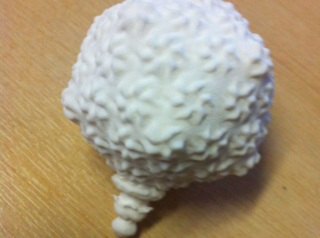
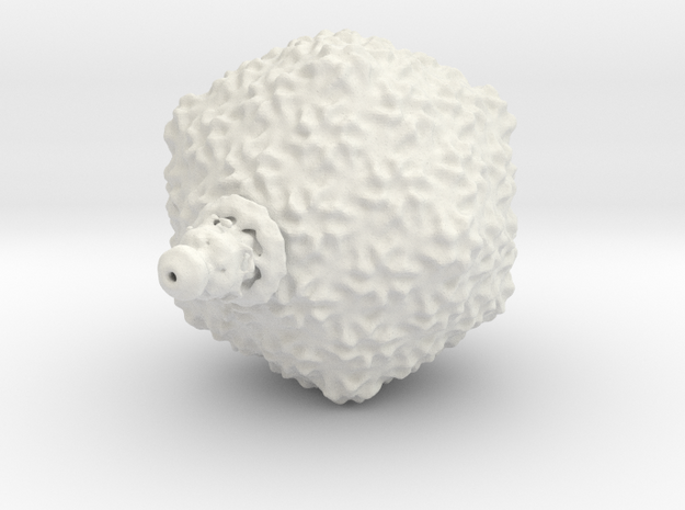
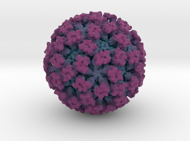
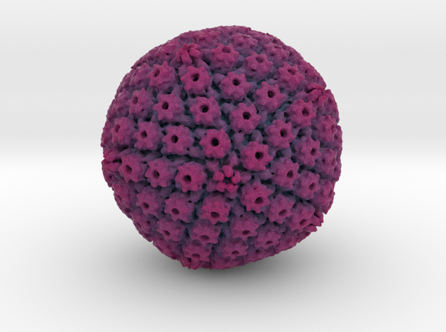
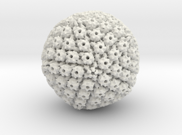
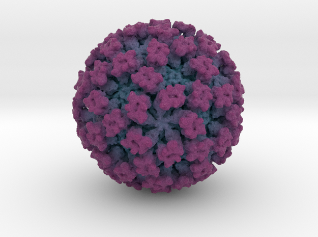
Viruses are the smallest pathogens to infect man. For reasons of genetic economy they assemble from a small number of building blocks, using symmetry. The resulting structures are both beautiful and intriguing.